Life of this generation people has become so simple with the help of the evolving technologies, no matter it is what in everything you can see some of the evolution when comparing to their past. This has become possible only with the help of the scientists, not only in the technology fields today you can see so many evolutions in the field of a science and it has got its name synthetic biology. The experts are considering synthetic biology it is a new frontier of science you might think about what the term synthetic biology means and their contribution to this world, to make you understand all those basics are coming in the below content.
What is synthetic biology?
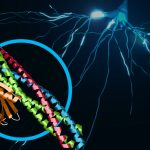
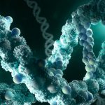
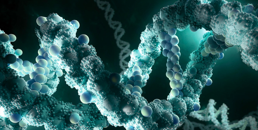
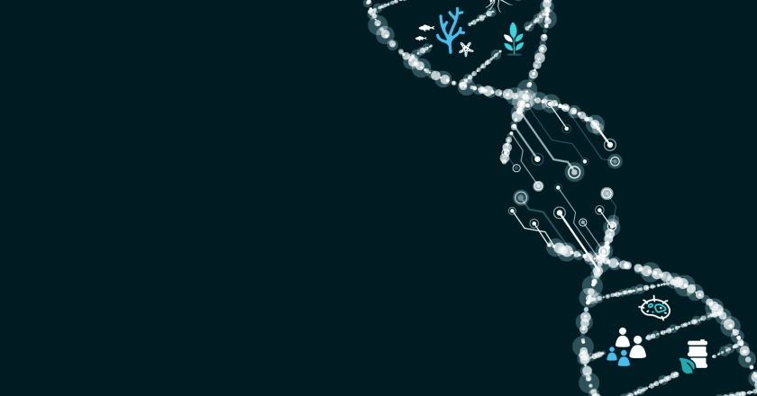
Generally, biology is a study that targets the living system and their environment and with the help of this biology, scientists have identifying each and everything single piece of information about the formation of the universe and the living creatures. Now, this has taken to the next step that is a trial to produce the things synthetically or artificially with understanding the basics of biology.
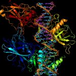
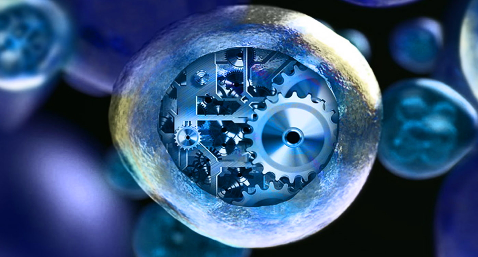
With the help of synthetic biology, scientists are trying to create a prokaryotic creature by providing all their requirements synthetically to check their growth and the genetic coding to understand whether it is possible to create living things synthetically. By understanding the organism genetically they are trying to incorporate the changes in their gene level with the help of the biotechnology and simulation tools they were started testing now they have achieved some levels in it successfully. This is said to be synthetic-engineering biology.
Not only the organism level now the research has reached to some extent and the scientists are also trying to develop the tissues, and organs in advance. The future trends in synthetic biology are keeping on increasing in recent years. Even most of them are ready to invest in projects related to synthetic biology because this is the field going to reach its peak in the next few years.
Through reading the above content you would have probably got the point why synthetic biology is considered to be the new frontier of science and also their contributions. As a human being you should know those things happening around you and to help synthetic biology explained above so read and update your knowledge on a relevant topic.